Clinical Impact of Volume Ultrasound for Musculoskeletal imaging
Introduction
Three-dimensional multi-planar ultrasound in combination with high-frequency transducers improves the understanding of complex anatomical relationships, assists in surgical planning of tendon and muscle tears, and measures the true volume of soft tissue masses. Volume Ultrasound is an important, non-invasive adjunct to two-dimensional (2D) imaging for musculoskeletal applications.
The Volume Ultrasound toolset includes Virtual Organ Computer-aided Analysis (VOCAL), which is a semi-automated measurement tool that is especially useful in calculating the volume of complex lesions. The volume is automatically calculated by the trackball tracing of a region of interest. By means of a step-by-step approach, VOCAL calculation builds its accuracy by adjusting the measurement to the shape of the target structure. Assumptions about the object's shape are no longer necessary.
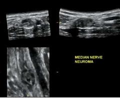
Soft tissue masses are easily identified and quickly diagnosed with Volume Ultrasound. The 4D16L volume transducer displays the anatomy in real time multi-planar mode. The coronal plane, unavailable with conventional ultrasound imaging, enables visualization of a mass and its relationship to surrounding vital structures in a fascinating new imaging plane. A surface rendering can also be created, allowing better characterization of the capsule of a mass.
The following cases demonstrate the clinical impact of Volume Ultrasound on the LOGIQ®9 ultrasound system (GE Healthcare, Wauwatosa, WI) for musculoskeletal imaging in the Henry Ford diagnostic radiology clinic.
Author:
Marnix T. van Holsbeeck, M.D.
Henry Ford Hospital, Detroit, MI