My Trainer
LOGIQ S8 XDclear 2.0
B-Flow
-

 Digitally encoded technology; using digital codes to enhance weak signals from small particulate reflectors and suppress signals from strong reflectors (tissue). Coded excitation and tissue equalization are key to B-Flow. Activation Created by the B-Mode processor. B-Flow is located on the keyboard next to the B-Mode button. B-Flow gain is controlled by turning the B-Mode gain button.
 -

Limits of B-Mode and Color Doppler
• B-Mode blood flow is displayed as black
• Color Doppler
a. Lack of true hemodynamics representation
b. Color writing on bright interfaces
c. Color outside vessel wall, over gain and high threshold
d. Inferior spatial resolution compared to B-Mode
e. ROI angle dependency.
-

Clinical Advantages of B-Flow
• Visualize both high and low velocity flow simultaneously
• No ROI to generate image •True hemodynamics, no overwriting on vessel wall
• Helps enhance spatial resolution of blood flow and less angle dependence than color flow
Examples High grade stenosis, Soft plaque or plaque ulcerations, Early thrombus, Organ perfusion such as Renal or placenta
-
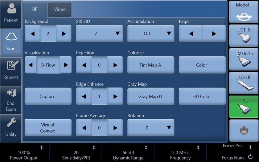
 Background Turn background off- 0 to suppress tissue signal allowing enhanced visualization of small vessels. Use 1-3 to determine the amount of background tissue to display to help identify surrounding landmarks in the image. Visualization B-Flow- Displays only B-Flow image Dual- Displays B-Mode and B-Flow images simultaneously using dual screen. Hybrid- Displays the B-Flow image over the B-Mode image using Hybrid Map. Press L and R at the same time to switch between Visualization values. The switch sequence can be selected in Utility -> Imaging -> BF -> “L/R Button Sequence”.
 -
 PRI/ Sensitivity For sensitivity to slow flow and small vessel identification, increase the PRI. Decreasing the PRI is useful in high flow conditions and helps minimize flash. The balance will vary based on probe and application used. Accumulation Use in slow flow states to help increase the time of optimal vessel visualization. During live scanning choose from the dropdown the fraction of a second the image will accumulate.
 -
Frame Average Increase frame average to help with smoother flow appearance.
If accumulation is used, frame average will be ignored.
Frequency Increase the frequency to enhance the detail.
Decrease the frequency for additional penetration.
Dynamic Range Increase dB level when less contrast is desired for a softer image.
To enhance the appearance of the flow, decrease dB level. A decrease in dB level increases contrast.
-

SRI HD To enhance the flow profile, increase SRI.
This can be done in post-processing images if desired.
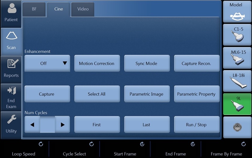
Cine Capture/Capture Cine Capture allows the user to represent the dynamic flow situation in the vasculature with a single still image.
Adjustments can be made to modify the start and end frames used in the process.
Select from the touch panel, a “c” will appear in the image parameter list in place of frame average.